massage and bodywork professionals
a community of practitioners
Neuroanatomy
The brain, Lesson 1
Objectives
By the end of this lesson, you will be able to do the following:
- distinguish between the prosencephalon, the mesencephalon, and the rhombencephalon;
- describe what is meant by “the reptilian brain” and “the mammalian brain”;
- distinguish between the telencephalon and the diencephalon.
Why you might be interested in knowing this: to orient yourself for more in-depth upcoming discussions of specific structures in specific areas of the brain.
Introduction
To understand where we are now, it is often very useful to know where we have been. In order to understand where our brains have been, we will look at both our embryological and our evolutionary histories.
The adult human brain is composed of gelatinous tissue, weighing about three pounds and containing about 100 billion neurons (nerve cells) and neuroglia (cells and tissues that support, protect, and nourish neurons). If you unwrinkled your brain and spread it out flat (not recommended, as that will void the original warranty), it would be about the size of a regular pillowcase; the wrinkles and folds are how it packs so many neurons and glia into such a small space as the skull can contain.
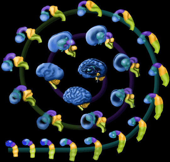
Embryologically, the brain is generated from an ectoderm tube that develops three divisions: the forebrain (prosencephalon, in red below), the midbrain (mesencephalon, in blue), and the hindbrain (rhombencephalon, in green).
First, what do we mean by an ectoderm tube? This is an outline of what the brain of an embryo looks like—it’s really a hollow tube, shaped like this:
Source: http://en.wikipedia.org/wiki/File:EmbryonicBrain.svg
You’re looking at it from the front or back in the outline. Now imagine rotating that outline 90 degrees to the side. That is the view that you have in this imaging study. You can start at the bottom left and follow the spiral up and around to the center to see how the embryonic tube develops and folds into a recognizable human brain. What do you think the color changes of the spiral (from teal to olive green to purple mean?

Source: http://www.visembryo.com/baby/NewsArchive31.html
The tube’s formed from ectoderm, which is the outer of the three layers of the vertebrate (= animals with backbones, like dogs, crocodiles, and us, and unlike insects, starfish, and like animals) embryo (as you might expect from the name “ectoderm”, the other two layers are called “mesoderm” and “endoderm”). This means that the brain develops from the same layer of the embryo that tooth enamel and the outer layer of skin come from.
The names “prosencephalon” and “mesencephalon” are easy to remember, since they carry their English elements “forebrain” and “midbrain”, but the source of the name “rhombencephalon” is not quite so obvious. Remember how in geometry, a square is a special kind of the diamond-shaped form called a “rhombus”? And how we have muscles called “rhomboids” in our backs?
Rhombus:
Source: http://en.wikipedia.org/wiki/File:Rhombus.svg
Square:
Source: http://en.wikipedia.org/wiki/File:3-simplex_t0.svg
Rhomboids:
Source: http://en.wikipedia.org/wiki/Rhomboid_muscles
Keeping that diamond shape in mind, look back at the rhombencephalon in the very first picture, and you’ll see the form that gave the hindbrain its technical name.
The midbrain and the hindbrain
The model of the triune brain, where the structures corresponded to different kinds of animals--the "lower" parts corresponding to the reptiles, and the "higher" parts corresponding to the earlier mammals and later mammals, is now regarded as too oversimplified to be taken totally at face value. As a model to start recognizing structures and finding our way around brains, it can be helpful. Just remember that it is not fully accurate, and don't take it too literally.
These two parts, midbrain and hindbrain, are often referred to as the most “primitive” parts of the brain, or as the “reptilian brain” in the triune model. They contain the basic neural machinery for reproduction and survival. When we use our reflexes, our survival instincts, when we feel territorial, want to find a mate, or eat—these are all times when our midbrain and hindbrain are active.
Some parts work on their own, without our even having to think about them—we don’t actually decide to have our hearts beat, to breathe, to flush when we get angry or hot. And behaviors such as aggression originate in this part of the brain as well.
The embryonic hindbrain eventually becomes the adult brain structures we call the pons, the cerebellum, and the medulla oblongata. We’ll talk in more detail about these structures later on. For now, just notice their shape--anatomists have compared them to a hummingbird and flower, as in the picture directly below.

Source: http://streetanatomy.com/2009/01/30/anatomic-fashion-friday-bird-br...
Can you see the hummingbird and flower in this picture (it's a little more abstract in this brain than in the previous one)?

Source: http://streetanatomy.com/2009/01/30/anatomic-fashion-friday-bird-br...
The forebrain
The forebrain is divided into two regions, the diencephalon in its rear part, and the telencephalon in its front. My anatomy teacher Naomi called the diencephalon “almost like the heart of your brain”—it’s the part often called “the mammalian brain”, concerned with emotional responses to stimuli, where emotions and memories are generated. Initial memories of smells, and social behaviours such as nest-building and nurturing of the young are also processed in the diencephalon (shown in blue, green, and yellow in this picture). (Since we've already decided to use the triune brain model more as a reminder than as a literal guide, we won't get into the fine differences between the paleomammalian [old mammals] and neomammalian [new mammals] structures here.)

Source: http://www.csuchico.edu/~pmccaffrey/syllabi/CMSD%20320/362unit5.html
The telencephalon (“end brain”) is the very frontmost part of the brain. It grows over and folds around the more primitive parts of the brain, and becomes the cerebrum (the two cerebral hemispheres). If you go back and follow that spiral picture of the embryonic brain, you can see how that develops--the structures at the front get bigger and bigger, then fold back over the other parts, and finally develop grooves and wrinkles.

Source: http://www.deryckthake.com/lecture-4-functional-neuroanatomy/
The cortex (its surface, from the Latin word for the bark of a tree) is where all of the primary sensory and motor regions are located. This is also where you find most of our advanced centers for thinking (the neocortex).
Questions to consider:

Source: http://sites.google.com/site/jwhidd/RatBrain2.jpg
The picture above is a rat brain. You are looking at it from the top of the rat’s head; if the head were still there, you would see that it is looking to the left of the picture.
- What do you notice about the texture of the rat’s brain?
- What does your observation have to do with the fact that there are no really great mathematicians who were rats?
- The two large projections at the left are structures called the olfactory bulbs. Below is a picture of the corresponding region in a human, except it’s from a different viewpoint than the rat picture is. This one is from the side, looking at the right half of the brain, with the left half sliced away. You can see the olfactory bulb inside the rectangle; it looks like a toothbrush (where the bristles are actually nerve endings). Let’s assume that the more important something is in an animal’s life is, the higher proportion of size, compared to the rest of the brain, is dedicated to, or invested in, the structures that support that function. Then what would you think rats are better at than humans are?

Source: http://www.worsleyschool.net/science/files/nose/page.html
At this point, Objectives 1, 2, and 3 should be comfortable for you. We'll build on these structures, and start to talk about functions in more detail in the next part.
Views: 1506
© 2026 Created by ABMP.
Powered by
![]()